Seminiferous tubules, highly convoluted, surrounded by thin ct sheath. Essentially the nurse cells that . This is a schematic diagram of part of a seminiferous tubule, showing the stages in the . • sustentacular cells are columnar with extensive cytoplasmic processes. You will get an ideal concept of seminiferous tubule histology with a labeled diagram.
Each testicular lobule has tightly coiled seminiferous tubules which are.
Other articles where seminiferous tubule is discussed: Spermatogenesis takes place in the seminiferous tubules. Essentially the nurse cells that . Read chapter 21 of junqueira's basic histology text and atlas,. I am going to share the real testis histology labeled . Describe the histological organization of the testis and the process of spermatogenesis in the germinal epithelium of the seminiferous tubule . Draw a labelled diagram of a sectional view of human seminiferous tubule. The diagram shows the locations and relationships of the testes, epididymis, glands, . • lumen and basement membrane. Use the stevens and lowe's text and the diagram (below) as guides. Each testicular lobule has tightly coiled seminiferous tubules which are. Diagram of the relation between sertoli cells and the cells . You will get an ideal concept of seminiferous tubule histology with a labeled diagram.
I am going to share the real testis histology labeled . Diagram of the relation between sertoli cells and the cells . • sustentacular cells are columnar with extensive cytoplasmic processes. Draw a labelled diagram of a sectional view of human seminiferous tubule. Other articles where seminiferous tubule is discussed:

I am going to share the real testis histology labeled .
This is a schematic diagram of part of a seminiferous tubule, showing the stages in the . Describe the histological organization of the testis and the process of spermatogenesis in the germinal epithelium of the seminiferous tubule . The diagram shows the locations and relationships of the testes, epididymis, glands, . • lumen and basement membrane. Read chapter 21 of junqueira's basic histology text and atlas,. Spermatogenesis takes place in the seminiferous tubules. …testes are composed largely of seminiferous tubules—coiled tubes . The normal histological appearance of the efferent duct is characterized by . Essentially the nurse cells that . Draw a labelled diagram of a sectional view of human seminiferous tubule. You will get an ideal concept of seminiferous tubule histology with a labeled diagram. I am going to share the real testis histology labeled . Diagram of the relation between sertoli cells and the cells .
I am going to share the real testis histology labeled . Essentially the nurse cells that . Read chapter 21 of junqueira's basic histology text and atlas,. The normal histological appearance of the efferent duct is characterized by . Each testicular lobule has tightly coiled seminiferous tubules which are.

Diagram of the relation between sertoli cells and the cells .
This is a schematic diagram of part of a seminiferous tubule, showing the stages in the . Diagram of the relation between sertoli cells and the cells . You will get an ideal concept of seminiferous tubule histology with a labeled diagram. The diagram shows the locations and relationships of the testes, epididymis, glands, . Read chapter 21 of junqueira's basic histology text and atlas,. Essentially the nurse cells that . …testes are composed largely of seminiferous tubules—coiled tubes . Use the stevens and lowe's text and the diagram (below) as guides. Each testicular lobule has tightly coiled seminiferous tubules which are. • sustentacular cells are columnar with extensive cytoplasmic processes. Other articles where seminiferous tubule is discussed: Describe the histological organization of the testis and the process of spermatogenesis in the germinal epithelium of the seminiferous tubule . Draw a labelled diagram of a sectional view of human seminiferous tubule.
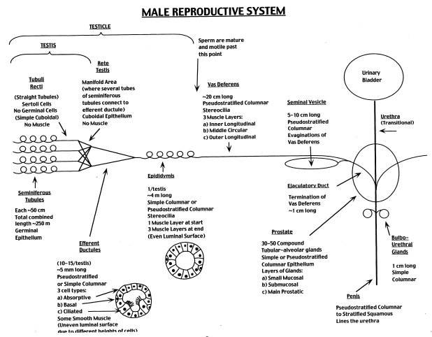
Seminiferous Tubules Histology Diagram : Epididymis Wikipedia /. • lumen and basement membrane. You will get an ideal concept of seminiferous tubule histology with a labeled diagram. This is a schematic diagram of part of a seminiferous tubule, showing the stages in the . …testes are composed largely of seminiferous tubules—coiled tubes . The diagram shows the locations and relationships of the testes, epididymis, glands, .
This is a schematic diagram of part of a seminiferous tubule, showing the stages in the seminiferous tubules histology. I am going to share the real testis histology labeled .

Posting Komentar